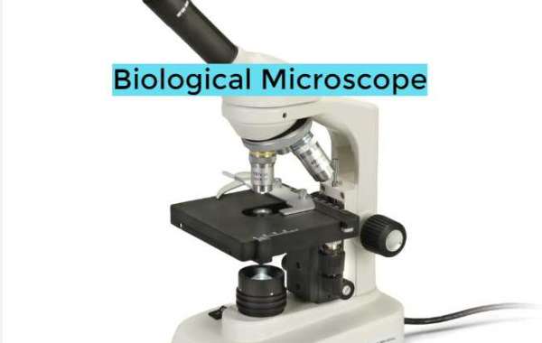
When it comes to the study and practice of pathology, Biological Microscope has evolved into tools that are absolutely necessary ever since their invention several centuries ago. Through the use of microscopes, pathologists are able to identify diagnostic microscopic changes and gain an understanding of disease processes at the fundamental levels of biology. This is accomplished by magnifying cellular and subcellular structures beyond the normal visual acuity. The purpose of this article is to investigate the ways in which the modern biological microscope and the associated techniques are the foundation for the numerous applications of pathology in the anatomical, clinical, and research fields.

Applications Derived from Anatomical Pathology
When attempting to diagnose disease states, anatomical pathologists examine tissues that have been removed or biopsied. Histological sections stained with hematoxylin and eosin (HE) and prepared from formalin-fixed paraffin embedded (FFPE) specimens are utilized in surgical pathology for the purpose of identifying abnormal cellular and subcellular morphology that is indicative of particular conditions. Architectural disorganization, inflammatory infiltrates, mitotic figures, pathological accumulations, and other microscopic indicators of cancer, infection, or injury/degeneration can be seen through the use of Biological Microscope. Intraoperatively evaluating surgical margins can be accomplished through the use of frozen section analysis, which involves rapid freezing and staining. The use of microscopy allows for precise diagnosis and directs subsequent treatment.
The functions of clinical pathology include the examination of body fluids, secretions, and excreta in order to identify the presence of diagnostic clues. An examination of thick and thin blood smears under a microscope allows for the morphological characterization of blood cells as well as the detection of parasitemia. In the presence of polarized light, cellular casts, crystals, and microorganisms can be identified through the analysis of urine sediment. It is a biological microscope. The process of isolating and identifying pathogenic microorganisms through the use of phase contrast microscopy is accomplished by microbiological cultures through the utilization of specialized culture media and a variety of incubation atmospheres. In order to identify infectious or cancerous cells, microscopy-guided analyses are performed on a variety of samples, including sputum, bronchial washings, synovial fluids, and other samples. Using brightfield microscopy, quantitative buffy coat testing is able to detect the presence of malarial parasites in blood in a very short amount of time. The use of microscopy continues to be a fundamental method in clinical pathology.
Various Applications of Forensic Pathology
Through the use of postmortem examinations and the microscopic identification of trace evidence, forensic pathologists contribute to the process of legal investigations. When preserved tissue sections, bone slivers, or swabs are examined using microscopy, the goal is to identify any projectiles, foreign debris, or pathogens that may indicate the cause and manner of death. When attempting to solve violent crimes or identify victims, comparisons to crime scene samples can be helpful. Additionally, fingernail scrapings, hairs, fibers, and documents that are under investigation are examined using microscopy in order to look for potential associative trace evidence. Compound Biological Microscope Suppliers are utilized on a regular basis as indispensable analytical tools in fields such as odontology, toxicology, and firearms examination.
Instructions for Histopathology
In addition to performing a general examination of HE stained slides, histopathologists conduct specialized staining procedures and microscopy examinations. Using trichrome stains, collagen fibers that are helpful in determining fibrosis can be highlighted. The acidic mucins that are secreted by goblet cells and glands can be identified by alcian blue. Staining with periodic acid-Schiff allows for the visualization of macromolecules in tissues that contain carbohydrates and serves as a diagnostic tool for fungal infections. Under fluorescence or confocal microscopy, immunohistochemistry makes use of enzyme- or fluorescent-labeled antibodies that are directed toward cellular antigens of interest. Specific DNA and RNA sequences in cells and tissues can be identified through the process of in situ hybridization. Examining organelle ultrastructure, viral particles, or amyloid fibrils can be accomplished with electron microscopy, which offers a resolution of nanometers. Diagnostic insights can be expanded through the use of complementary microscopic modalities.
The Applications of Research
Not only do microscopes serve clinical and diagnostic purposes, but they also contribute to the advancement of research in the life sciences. By conducting experiments under the guidance of microscopy, cellular and molecular pathologists investigate the mechanisms underlying disease. Live cell imaging is a technique that investigates the dynamic processes that occur within microorganisms and cells that are either genetically modified or fluorescently labeled. In order to bridge resolutions and shed light on subcellular changes, correlational light-electron microscopy is utilized. The screening of tumors for prognostic factors or therapeutic response predictors is accomplished through the use of tissue microarrays that have been stained with clinically validated biomarkers. Through the use of microscopic quantification, parameters that are helpful for drug discovery or precision medicine initiatives can be extracted. All things considered, the Biological Microscope Supplier continues to be an indispensable research instrument that propels the advancement of medical technology.
Particularly Tailored Applications
Skin biopsy specimens are examined by dermatopathologists to determine whether or not they contain cancers, inflammatory diseases, or genetic keratinization disorders. Ophthalmic pathology is the study of identifying abnormalities in ocular tissue as well as infectious agents that have an effect on vision. The field of neuropathology examines the changes in neural tissue that are the root cause of neurological disorders. In dental pathology, both hard and soft oral tissue pathology are diagnosed and treated. For the purpose of investigating zoonotic, agricultural, and companion animal diseases, veterinary pathology employs techniques that are similar to one another. Through the use of microscopy, comparative methodologies can be standardized across a wide variety of specialized pathology domains.


